
Accommodation
In many ways, the eye functions rather like a camera and accommodation refers to the ability to see well close up as well as in the distance.
Loss of accommodation is also called presbyopia. This is a bit like losing the ' zoom' function on a camera. People over the age of 45 (and sometimes earlier) who may previously have had good vision, find they need spectacles to read small print. This is the onset of presbyopia or loss of accommodation.

Amblyopia
Also known as "lazy eye."
Decreased vision in one or both eyes without detectable anatomic damage in the eye or visual pathways. spectacles or contact lenses do NOT usually solve this problem.
During the first 5-8 years of life, a child's vision is developing. If during that time one eye is misaligned ( strabismus or squint) or obstructed, the brain will suppress the image and concentrate on the better eye. The weaker eye, therefore, does not develop properly. The condition is not always obvious, but any if left untreated beyond the age of 8, will continue into adult life.
Causes of Amblyopia
An obstructed view may be due to cataract. Strabismus or squint.
Anisometropia
Where the focusing power is unequal leaving one eye that does not focus well, usually due to long or short-sightedness.
Treatments
Untreated amblyopia can lead to permanent visual problems and lack of depth perception. Also, if a person's "good eye" develops a condition or is injured later in life, the amblyopic eye will not see well enough to provide adequate vision.
Correcting the vision in the amblyopic eye will depend on the cause. If amblyopia is due to a cataract, this should be removed urgently.
If it is due to strabismus it can be treated with a combination of eye drops, glasses, patching and/or eye muscle surgery.
Anisometropic amblyopia is treated with glasses to correct the focusing problem.
Astigmatism
If the curvature of the front of the eye is uneven, the light will refract unequally and may result in blurred vision.
Many people are unhindered by a little astigmatism although having more than 1 dioptre especially if it is combined with other refractive conditions such as myopia (short-sightedness) or long-sightedness ( hyperopia) may require correction.
Astigmatism can be corrected by glasses and sometimes by contact lenses. It can also be corrected with laser surgery or by surgical techniques during cataract or lens replacement surgery.

Blepharitis
Blepharitis is a common and persistent inflammation of the eyelids. Symptoms include irritation, itching and occasionally, red-eye.
The bacteria that live on everyone's skin sometimes live within the hair follicles at the base of the lashes where they can cause an overproduction of oil, resulting in a scaley itchy residue on the lash margins.
Some people develop a further reaction, resulting in inflammation of the eye tissue and cornea (the transparent part of the front of the eye).
Treatment of Blepharitis
Blepharitis can be controlled with a few simple daily measures: At least twice a day, wet a clean flannel with comfortably warm water, wring it out, and place over the closed eyelids for a minute. As it cools, re-wet it, repeating several times. This will soften and loosen scales and debris. More importantly, it helps prevent the oily secretions from hardening and forming an inflamed lump, also known as a chalazion.
With a moist cotton bud, or commercial lint-free pad, gently scrub the base of the lashes about 15 seconds per lid.
If an antibiotic ointment has been prescribed, apply a dot at the base of the lashes (usually at bedtime), using your fingertip. The above measures will minimise the symptoms but additional medications may be needed to control blepharitis and its symptoms:
Artificial tears may be used to relieve symptoms of dry eye. (Preservative free ones are available, these can be bought from a pharmacy). Steroid eye drops may be used short-term to decrease inflammation. Antibiotic ointment may be used to decrease the bacterial content of the eyelids.
Antibiotic tablets may be used to decrease the oil production from the meibomian glands.
Blocked Tear Duct

The tear film protects the eye from infection and allows clear vision. It constantly replenishes itself, washing across the front of the eye, entering the inner corner of the eye, and passing downward into the tear sac and through a vertical passage (the tear duct) to the back of the nose.
Babies often have tear duct blockages which can usually be cleared with daily massaging of the side of the nose.
In adults, if the tear duct does not open on its own or with the aid of massage, it may be necessary for your ophthalmologist to unblock it using a small probe. This is a minor procedure lasting no more than twenty minutes, which resolves the problem, 90% of the time, however sometimes a second probing is needed.
Symptoms are usually when tears flow onto the cheek. If the tears lead to crusting, there may be an infection.
Persistent problems should be seen by an ophthalmologist to ensure there is no physical blockage of the duct.
Chalazion
Inflamed lump in one of the oil-producing glands (in the eyelid margin) of either upper or lower lid. Inflammation usually subsides but may need surgical removal. Sometimes called an internal hordeolum. A chalazion may be confused with a ' STYE' which is inflammation of an eyelash follicle.
Treatment
Chalazion may be treated with any one or a combination of antibiotic or steroid drops or injections; warm compresses for 5 to 10 minutes, 3 or 4 times a day; gentle massage to express the glandular secretions; or surgical drainage. Chalazions usually respond well to treatment, although some people are prone to recurrences and may require continuing medication. If a chalazion recurs in the same place, a biopsy may be suggested, to rule out a more serious problem. Occasionally, a chalazion can cause the entire eyelid to swell suddenly.
Conjunctivitis
The conjunctiva is the transparent membrane covering the outer surface of the eyeball, sometimes called 'the window of the eye'
Conjunctivitis is also known as "pink eye". It is inflammation of the conjunctiva. Characterised by discharge,
grittiness, redness and swelling.
Usually viral in origin, but may be bacterial or allergic; may be contagious.
Sometimes newborn babies develop conjunctivitis as a result of the spread of infection from the mother during birth. Any parent who suspects their newborn baby has conjunctivitis should contact their GP, health visitor or midwife.
Conjunctivitis can also be caused by an allergy. Common allergies are pollen ( hay fever) and dust mites as well as allergies to pets and mould.
Treatment
Irritation in the eyes can be partially relieved by bathing them with preparations available over the counter from a pharmacist. These often contain witch hazel, which is thought to have soothing properties. Contact lens wearers should remove their contact lenses.
Avoid rubbing your eyes as this makes the inflammation worse. To avoid spreading the condition from one eye to another or to other people( in bacterial conjunctivitis) you should not touch the affected eye. Any towels used to dry the affected eye after washing should not come into contact with the other eye or be used by other people.
The most important treatment for allergic conjunctivitis is to avoid the substance that causes the allergy.
Bacterial conjunctivitis can be treated with antibiotic eye drops or ointment, which can only be obtained with a prescription from your doctor.
Allergic conjunctivitis is treated with eye drops that contain sodium cromoglicate, which can be obtained with a prescription from your doctor, or bought over-the-counter from a pharmacist (eg Optrex Allergy, Opticrom Allergy). Always follow the instructions that come with the medicine. Generally, the drops should be applied to both eyes four times a day. It is important that eye drops are applied regularly throughout the hay fever season, regardless of how severe the symptoms are on any particular day.
Viral conjunctivitis does not need specific medical treatment and will clear up on its own in two to three weeks.
Corneal Abrasions & Erosion
Corneal Abrasion
The cornea is the clear front window of the eye. A corneal abrasion is an injury to the surface layer of the cornea. Abrasions are painful.
The corneal surface usually heals within a day or two, but the eye may be very uncomfortable while it is healing. Tearing, light sensitivity and the feeling that something is in the eye - "foreign body sensation" - will accompany even a small abrasion.
A common treatment is to patch the scratched eye. Another common treatment is repeated application of ointment to the eye, which forms a soothing layer between the inner eyelid and the abrasion. Antibiotics are often used because of the small risk of infection. Sometimes a drop is used to dilate the pupil to help with pain associated with light sensitivity.
Even after the surface has healed, the cornea may still be sensitive to wind and dust. Often, additional lubrication is helpful, both during the day and at bedtime, until the sensitivity has disappeared. Some other diseases, such as dry eye or diabetes, may be slow healing.
What is a corneal erosion?
Corneal erosion is a spontaneous breakdown of the surface of the cornea. The symptoms are similar to a corneal abrasion: An erosion may occur when the eyes are dry or irritated.
How are corneal erosions treated?
Several treatments are used to alleviate the discomfort of erosions and to speed healing:
-
Lubricating drops and ointments;
-
Drops or ointments containing salt;
-
A special contact lens used to bandage the cornea;
-
Micro-puncture of the epithelium;
-
Removal of the damaged epithelium.
-
Recurrent corneal erosions can be stubborn and frustrating. Professor Aggarwal may be able to identify other contributing factors. Applying the appropriate drops or ointment as prescribed is often the key to ending the erosion cycle.

Eye

Eyelid Cancer
Cancer can occur within the eye as well in the tissues surrounding the eye. There are several forms of cancer from painless nodules to crusty areas, areas that are raised or even bleeding.
The most common cause of skin cancer is overexposure to sunlight and is more prevalent in fair-skinned people.
Types of eyelid skin cancer:
-
Basal cell carcinoma (this type tend not to spread to distant parts of the body)
-
Squamous cell carcinoma (this type tend not to spread to distant parts of the body)
-
Sebaceous gland carcinoma (This is a more serious type of skin cancer because it may spread to other parts of the body)
-
Malignant melanoma (This is a more serious type of skin cancer because it may spread to other parts of the body)
-
Complete removal of the tumour is necessary to minimise the possibility that it may recur. Reconstructive surgery may also be necessary.
Glaucoma
Glaucoma is a condition that affects around 2% of people over the age of 40 in the UK. It causes damage to the optic nerve, which can lead to vision loss and potentially blindness if left untreated. The most common cause is increased pressure inside the eye, although some people may develop optic nerve damage even when eye pressure is normal.
There are two main types of glaucoma. Chronic (open-angle) glaucoma develops gradually over time and is usually painless, while acute (closed-angle) glaucoma occurs suddenly, can be painful, and requires immediate treatment to prevent permanent vision damage.
Glaucoma can often be detected during a routine eye examination, making regular eye tests important, particularly for individuals with a family history of the condition.

Herpes Simplex of the Eye
Herpes simplex is a very common virus affecting the skin, mucus membranes, nervous system, and the eye. There are two types of herpes simplex.
Type I causes cold sores or fever blisters and may involve the eye.
Type II is sexually transmitted and rarely causes ocular problems.
Nearly everyone is exposed to the virus during childhood. The initial infection is usually mild, causing only a sore throat or mouth. After exposure, herpes simplex usually lies dormant in the nerve that supplies the eye and skin.
Later on, the virus may be reactivated by stress, heat, running a fever, sunlight, hormonal changes, trauma, or certain medications. It is more likely to recur in people who have diseases that suppress their immune system. In some cases, the recurrence is triggered repeatedly and becomes a chronic problem.
When the eye is involved, herpes simplex typically affects the eyelids, conjunctiva, and cornea. Keratitis (swelling caused by the infection), a problem affecting the cornea, is often the first sign of the disease in eyes. In some cases, the infection extends to the middle layers of the cornea, increasing the possibility of permanent scarring. Some patients develop uveitis, an inflammatory condition that affects other eye tissues.
Signs and Symptoms
Pain, Red Eye, Light Sensitivity, Irritation, possible visual problems
Treatment
Usually, drops or antiviral medication will resolve the situation. An unresolved ongoing problem can lead to permanent corneal scarring which may require a corneal graft.
Hyperopia - Long Sighted
People who are long-sighted may be able to see well at distance but often can't see well close up. Those with mild hyperopia may be able to manage without vision correction, whilst those with higher prescriptions or who are older may find it difficult to focus at any distance.
An eye that sees well without spectacles is the right length and therefore light falls onto the retina where the image is processed by the brain to form a clear picture.
A long-sighted eye is usually too small or the cornea is too flat. If the lens cannot compensate (presbyopia) the image focuses behind the retina, rather than on the retina, causing a blurred image.
A plus correction is needed to correct the problem. If you have a +2.00D lens prescription for example, then you are long or farsighted.
Permanent solutions to the problem are also available, including LASIK and LASEK. Excellent results can also be obtained with Refractive Lens Exchange (also known as Clear Lens ExChange).

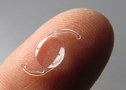
IOL - Intraocular Lens
This is a synthetic lens that is implanted in the eye to replace the natural lens. lenses have different focal strengths (called dioptres) and the initial measurements taken of the eye, determine which size is best.
There are many different types and styles, usually, they are silicone or acrylic foldable lenses (which can vary in design). They can be fixed or single focus (monofocal) or variable focus (bifocal, trifocal, accommodative or multifocal).
Some lenses are specially made to deal with astigmatism as well as short or long sight and these are called 'toric' lenses. Lenses are usually round in shape and are approximately 5-6mm across the optic or central portion. They are inserted into the eye via a tiny incision (less than 2mm).
To ensure they remain stable within the eye, they have stabilising arms called haptics. In a one-piece lens, the optic and haptic are as one, whilst they are joined together in the 3 piece version.
The intraocular lens is positioned behind the iris it is not usually visible from the front and would expect to remain in place for a lifetime.
Myopia - Short-sight
An eye that sees well without spectacles is the right length and therefore light falls onto the retina where the image is processed by the brain to form a clear picture.
A short-sighted eye is usually too long and the image focusses in front of the retina, rather than on the retina, causing a blurred image.
A minus correction is needed to correct the problem. If you have a -2.00D spectacle prescription for example, then you are short-sighted.
Permanent solutions to the problem are also available, including LASIK and LASEK.
Phakic IOL's are also available for people who are very short-sighted.
Myopia means short-sighted. People who are short-sighted can see objects close up but cannot see in the distance.
Many people cannot read or use a computer without spectacles because the distance at which they would focus without spectacles is too close for comfort.
Generally, short-sighted people would need spectacles for most things including driving and computer work, but some may be able to read or thread a needle without specs.
Nystagmus
Nystagmus is an uncontrolled movement of the eyes, usually from side to side, but sometimes the eyes swing up and down or even in a circular movement. Most people with nystagmus have reduced vision.
Babies can be born with the condition and this is called 'congenital nystagmus' whilst onset later in life is referred to as acquired nystagmus'. If a child is born with the condition it is not always a symptom of any other problem and many lead perfectly normal lives.
Late-onset of the condition can be linked to other conditions such as stroke, multiple sclerosis or a blow to the head. All patients suffering from this condition should be referred to an ophthalmologist.

Treatment
If the patient has other refractive errors such as long sight, short sight or astigmatism, these can e corrected with spectacles, although the nystagmus cannot be corrected. Some eye care specialists have developed programmes to help nystagmus patients control the problem.
Occasionally surgery may be undertaken to reduce uncontrolled movement. There are no real cures for the problem however sufferers do get used to dealing with it and it does not result in blindness.
For comprehensive information about nystagmus see 'The Nystagmus Network '
Refractive Error
Almost all eyes will have some refractive error. If they are of the ' higher-order' variety the brain will ignore these most of the time. Indeed they may not be evident until later in life when they are combined with other disorders such as cataract or presbyopia.
For ' lower order' variations, however, correction is often needed.
These include myopia or shortsightedness, hyperopia or long-sightedness, astigmatism and presbyopia.
Lower order refractive error can be detected at a routine eye examination.
Mr Aggarwal uses wavefront analysis to detect and treat higher-order aberrations during the treatment of lower-order variations, to provide the very best possible outcome.
